
Virumbrales-Muñoz Lab Home Page

Thank you to our funding sources:
- Diane Lindstrom Foundation
- University of Wisconsin Carbone Cancer Center
- University of Wisconsin Centennial Scholars
- Meriter Foundation
- American Cancer Society
Research Summary:
- Biomimetic modeling of ovarian cancer initiation and factors promoting metastasis.
- Biomimetic modeling of cell-cell interactions and cues in the primary breast tumor.
- Biomimetic modeling of tumor angiogenesis (e.g., renal, ovarian).
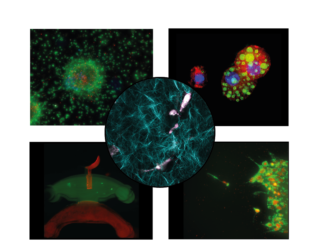
Figure caption: (Top left) Ovarian cancer spheroids being targeted by engineered cytotoxic immune cells (white/blue). Green and red show live and dead cells, respectively. (Top right) Differentiated adipocytes embedded in 3D showing lipid droplets (green), F-actin cytoskeleton (red), nuclei (blue). (Center) NADH autofluorescence (pink) in breast cancer fibroblasts embedded within a cyan-depicted collagen network. (Bottom left) Novel microfluidic device to study serous tubal intraepithelial carcinoma (STIC) lesions. (Bottom left) Ovarian cancer spheroids live tracked for mitochondrial organization (green) in respect to nuclei location (red).
Our research focuses on understanding the microenvironmental cues driving cancer progression and metastasis, with a specific focus on finding molecular targets to slow these processes. To this end, we generate physiologically relevant 3D models of tissue microenvironments using microfluidic technologies (i.e., small platforms handling volumes in the microscale). These technologies allow us to assemble different cell types in structures that resemble tissue organization, and cancer progression in these tissues. Specifically, we work on modeling the Fallopian tube and omentum for ovarian cancer metastasis, modeling the mammary duct for breast cancer, and the renal tubule for kidney cancer. We use a combination of molecular and functional techniques (e.g., microscopy with fluorescent reporters, bead-based ELISA) to characterize these models and evaluate the role of specific cues in driving cancer progression and metastasis. Model results are validated back to patient data with the ultimate goal of improving health outcomes.